Pervichny akusherskiy skrining (1 trimester):
First Trimester Screening During Pregnancy:
What is it?
• A routine ultrasound examination conducted in the first trimester of pregnancy.
• Typically scheduled between 11 and 14 weeks of gestation.
• The goal is to assess early fetal development and identify potential risks of genetic and congenital abnormalities.
Why is the first screening done?
• To confirm the viability of the fetus (presence of heartbeat and movement).
• To determine the exact gestational age.
• To assess the number of fetuses (singleton or multiple pregnancy).
• Early diagnosis of congenital defects and chromosomal abnormalities.
• To check the development of the fetus's internal organs and structures.
What can be seen on the ultrasound?
• The growth and size of the fetus in relation to the gestational age.
• The thickness of the nuchal translucency (NT) — an important marker for potential chromosomal abnormalities.
• The length of the nasal bone.
• The formation of the heart, stomach, bladder, limbs, and spine.
• The development of the placenta and the amount of amniotic fluid.
When is it scheduled?
• Routinely for all pregnant women between 11 and 14 weeks of gestation.
• Additionally, if there is an increased risk of genetic disorders or unfavorable heredity.
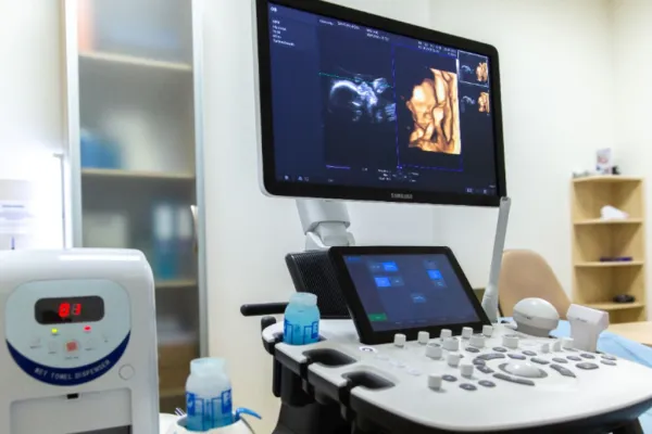
How is the procedure performed?
• Most commonly performed through the abdomen (transabdominally).
• In some cases, it may be conducted through the vagina (transvaginally) for a more precise image.
• A special gel is applied to the abdomen, the doctor moves the transducer, and the image of the fetus is displayed on the screen.
• The procedure lasts about 10-15 minutes.
What is it?
• A routine ultrasound examination conducted in the first trimester of pregnancy.
• Typically scheduled between 11 and 14 weeks of gestation.
• The goal is to assess early fetal development and identify potential risks of genetic and congenital abnormalities.
Why is the first screening done?
• To confirm the viability of the fetus (presence of heartbeat and movement).
• To determine the exact gestational age.
• To assess the number of fetuses (singleton or multiple pregnancy).
• Early diagnosis of congenital defects and chromosomal abnormalities.
• To check the development of the fetus's internal organs and structures.
What can be seen on the ultrasound?
• The growth and size of the fetus in relation to the gestational age.
• The thickness of the nuchal translucency (NT) — an important marker for potential chromosomal abnormalities.
• The length of the nasal bone.
• The formation of the heart, stomach, bladder, limbs, and spine.
• The development of the placenta and the amount of amniotic fluid.
When is it scheduled?
• Routinely for all pregnant women between 11 and 14 weeks of gestation.
• Additionally, if there is an increased risk of genetic disorders or unfavorable heredity.
How is the procedure performed?
• Most commonly performed through the abdomen (transabdominally).
• In some cases, it may be conducted through the vagina (transvaginally) for a more precise image.
• A special gel is applied to the abdomen, the doctor moves the transducer, and the image of the fetus is displayed on the screen.
• The procedure lasts about 10-15 minutes.


