Ultrasound Examination (Ultrasound) of the Lungs:
• A modern diagnostic method based on the use of ultrasound waves to assess the condition of the lungs and pleural cavity.
• A painless, safe, and radiation-free method.
• Can be performed repeatedly for dynamic observation.
Why Lung Ultrasound is Performed:
• To detect fluid in the pleural cavity (effusion, hydrothorax).
Why Lung Ultrasound is Performed:
• To detect fluid in the pleural cavity (effusion, hydrothorax).
• When inflammatory processes are suspected (pneumonia, pleurisy).
• To assess the condition of the pleura, diaphragm, and chest wall.
• To monitor the treatment of lung and pleural diseases.
• In emergency cases — for rapid diagnosis of chest injuries, pneumothorax.
What Can Be Seen During the Examination:
• Presence and volume of fluid in the pleural cavity.
• Areas of inflammation or consolidation in lung tissue.
• Condition of the pleura and diaphragm.
• Indirect signs of pneumothorax (air accumulation).
• Condition of the chest wall, tumor formations near the pleura.
When Lung Ultrasound is Prescribed:
• In cases of shortness of breath, cough, chest pain.
• When pneumonia, pleurisy, or other respiratory system diseases are suspected.
• To detect fluid or air in the pleural cavity.
• For monitoring after chest surgeries and injuries.
• When X-ray or CT is not possible (e. g. , in pregnant women).
Preparation for the Procedure:
• No special preparation is required.
• No need to restrict food or drink before the examination.
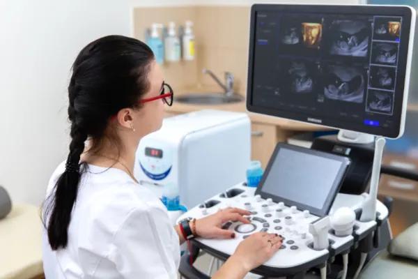
How the Procedure is Conducted:
• The patient sits or lies down, depending on their condition.
• The doctor applies a special gel to the skin of the chest.
• The sensor is moved across different areas of the chest, and images of the organs are displayed on the screen.
• On average, the examination takes 10–15 minutes.
• After the ultrasound, you can immediately return to your usual activities.
Вас може проконсультувати лікар вищої категорії, КМН, Перелигін І.В.