Ultrasound examination of the heart, or echocardiography, is a modern and safe method that allows real-time assessment of the heart's function, structure, and blood flow.
When it is prescribed:
• for shortness of breath, chest pain, or irregular heartbeats;
• for arrhythmias, high blood pressure, or swelling;
• to monitor the condition after a heart attack or heart surgery;
• for congenital and acquired heart defects;
• to evaluate heart function in athletes and patients with chronic diseases.
What the ultrasound shows:
• the size and shape of the heart chambers;
• the thickness of the walls and myocardial contractility;
• the condition of the valves and blood flow through them;
• the presence of defects, septal defects, clots, or tumors;
• pressure in the pulmonary artery and signs of heart failure.
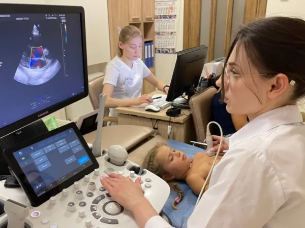
How the procedure is performed:
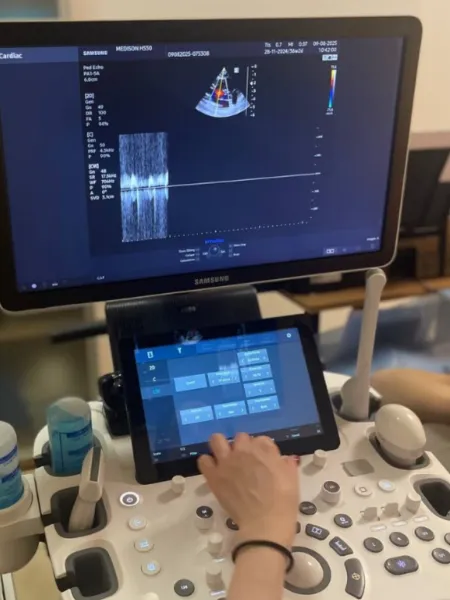
The patient lies on a couch on their back or left side. Gel is applied to the chest, and the doctor places an ultrasound probe to obtain an image of the heart on the screen. If necessary, a Doppler study is conducted to assess the speed and direction of blood flow. The procedure lasts 20–30 minutes and requires no preparation.
Advantages of the method:
• safe, painless, and radiation-free;
• allows early detection of diseases;
• provides the doctor with a complete live picture of heart function;
• can be repeated multiple times, including for treatment monitoring;
• suitable for children, adults, and pregnant women.
Вас может проконсультировать врач высшей категории, КМН, Перелыгин И.В.
Adult heart ultrasound (echocardiography) at the "Genesis Dnipro" clinic — accurate heart function diagnostics...
Подробнее
Speckle Tracking Echocardiography (STE) at the "Genesis Dnipro" clinic — innovative heart diagnostics.
Подробнее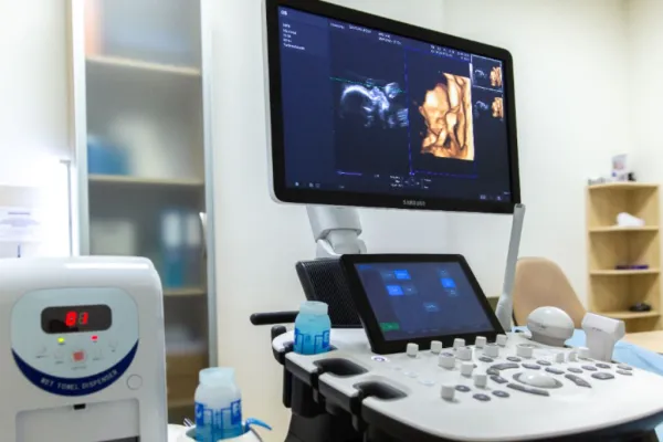
Fetal heart ultrasound (intrauterine) at the "Genesis Dnipro" clinic — an accurate assessment of the baby's he...
Подробнее


