Dopplerometry of the fetus
Dopplerometry during Pregnancy:
What is it?
• It is an ultrasound diagnostic method that allows for the assessment of blood flow in the vessels of the mother, placenta, and fetus.
• It is based on the Doppler effect, which is the change in frequency of ultrasound waves when reflected off moving particles (in this case, red blood cells).
• It is a safe and painless method that can be performed multiple times.
Why is Dopplerometry performed?
• To assess the delivery of oxygen and nutrients to the fetus through the placenta.
• To detect signs of hypoxia (oxygen deprivation of the baby).
• To diagnose intrauterine growth restriction of the fetus.
• To check the condition of the umbilical vessels, uterine arteries, and fetal brain vessels.
• To monitor the effectiveness of treatment in case of pregnancy complications.
What can be observed during the examination?
• The quality of blood flow between the mother, placenta, and fetus.
• Dysfunction of the placenta (placental insufficiency).
• Signs of hypoxia in the baby.
• Risk of fetal growth restriction.
When is it prescribed?
• Routinely, most often along with the third screening (30–34 weeks).
• Unscheduled, in cases of suspected hypoxia, oligohydramnios or polyhydramnios, fetal growth restriction, placental pathology, preeclampsia, or high blood pressure in the mother.
How is the procedure conducted?
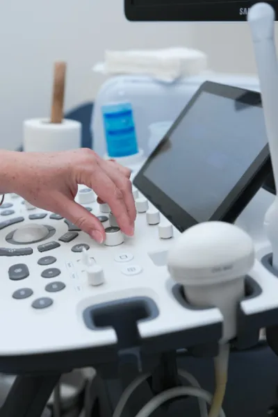
• It is performed similarly to a regular ultrasound: the patient lies on a couch, and the doctor moves a transducer over the abdomen.
• Graphs and color images of blood flow in the vessels are displayed on the screen.
• The procedure takes about 10 minutes.
• No preparation is required.
What is it?
• It is an ultrasound diagnostic method that allows for the assessment of blood flow in the vessels of the mother, placenta, and fetus.
• It is based on the Doppler effect, which is the change in frequency of ultrasound waves when reflected off moving particles (in this case, red blood cells).
• It is a safe and painless method that can be performed multiple times.
Why is Dopplerometry performed?
• To assess the delivery of oxygen and nutrients to the fetus through the placenta.
• To detect signs of hypoxia (oxygen deprivation of the baby).
• To diagnose intrauterine growth restriction of the fetus.
• To check the condition of the umbilical vessels, uterine arteries, and fetal brain vessels.
• To monitor the effectiveness of treatment in case of pregnancy complications.
What can be observed during the examination?
• The quality of blood flow between the mother, placenta, and fetus.
• Dysfunction of the placenta (placental insufficiency).
• Signs of hypoxia in the baby.
• Risk of fetal growth restriction.
When is it prescribed?
• Routinely, most often along with the third screening (30–34 weeks).
• Unscheduled, in cases of suspected hypoxia, oligohydramnios or polyhydramnios, fetal growth restriction, placental pathology, preeclampsia, or high blood pressure in the mother.
How is the procedure conducted?
• It is performed similarly to a regular ultrasound: the patient lies on a couch, and the doctor moves a transducer over the abdomen.
• Graphs and color images of blood flow in the vessels are displayed on the screen.
• The procedure takes about 10 minutes.
• No preparation is required.


