Ultrasound examination (US) of the mammary glands is a modern and safe diagnostic method that helps the doctor assess the condition of breast tissues. The procedure is painless, does not involve radiation, and is suitable for repeated use.
When it is prescribed:
• when lumps, pain, swelling, or nipple discharge appear;
• to clarify data from mammography or MRI;
• when cysts, nodules, or fibroadenomas are detected;
• to monitor previously identified formations;
• if inflammatory processes are suspected;
• for preventive purposes — especially for women under 40, pregnant, and breastfeeding women.
What the ultrasound shows:
• the structure of breast tissues;
• cysts, benign and malignant formations;
• signs of inflammation (mastitis, etc. );
• the condition of the milk ducts and axillary lymph nodes.
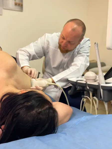
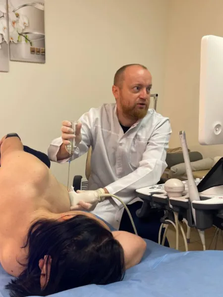
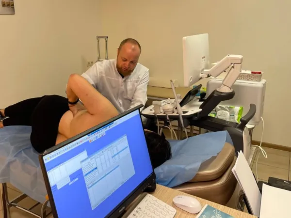
How the procedure is conducted:
The patient lies on her back, and a special gel is applied to the skin. The doctor moves the ultrasound probe over the surface of the breast and axillary areas, and the image is displayed on the screen. The examination takes 15–20 minutes, and no preparation is required.
Advantages of the method:
• painless and safe;
• no X-ray radiation is used;
• can be performed frequently, including during pregnancy and breastfeeding;
• suitable for preventive examinations;
• provides the doctor with detailed information about the condition of breast tissues.
Вас може проконсультувати лікар вищої категорії, КМН, Перелигін І.В.