What is an ultrasound?
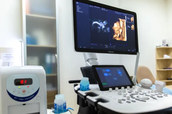
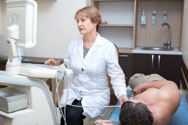
Ultrasound (US) is a safe, painless, and informative diagnostic method based on the use of ultrasonic waves. It allows doctors to see the condition of internal organs, tissues, and body systems in real-time.
Ultrasound is widely used in gynecology, obstetrics, urology, cardiology, endocrinology, surgery, and other medical fields.
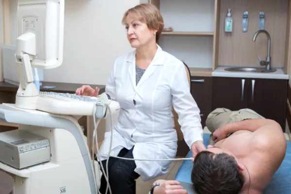
Ultrasound is widely used in gynecology, obstetrics, urology, cardiology, endocrinology, surgery, and other medical fields.
What is Doppler ultrasound?
Doppler ultrasound (or ultrasound with Doppler) is a type of ultrasound examination that assesses blood flow in vessels. This method allows doctors to determine:
- the speed and direction of blood flow;
- the condition of the vessel walls;
- the presence of narrowing or blockages;
- the quality of blood supply to organs and tissues.
In obstetrics, Doppler ultrasound is indispensable for monitoring blood circulation in the "mother-placenta-fetus" system, helping to timely identify risks of hypoxia or developmental delays in the child.
When are ultrasound and Doppler ultrasound recommended?
Ultrasound is recommended:
- during preventive check-ups;
- when there are complaints of pain or organ dysfunction;
- for monitoring chronic diseases;
- during pregnancy to assess the condition of the fetus and mother.
Doppler ultrasound is conducted when:
- there is suspicion of vascular pathologies (thrombosis, atherosclerosis, varicose veins);
- planning and during pregnancy;
- there are circulatory disorders in the brain, heart, kidneys;
- there is suspicion of hypertension, ischemia, diabetic angiopathy.
Advantages of the method:
- Safety: Ultrasound and Doppler have no radiation exposure and are suitable even for pregnant women and children.
- Informativeness: allows for the detection of pathology at an early stage.
- Speed: the examination takes 10–30 minutes.
- Accessibility: does not require complex preparation and is conducted on an outpatient basis.
How to prepare for an ultrasound?
Preparation depends on the area of examination:
- for abdominal organs — come on an empty stomach;
- for the pelvic area — sometimes a full bladder is required;
- for vascular studies — no special preparation is needed.
Before the procedure, the doctor always explains all recommendations in detail.
Why choose our clinic?
- state-of-the-art expert-class equipment;
- experienced ultrasound specialists and obstetricians-gynecologists;
- comfortable examination conditions;
- the possibility to immediately receive a specialist consultation and treatment plan.
Вас може проконсультувати лікар вищої категорії, КМН, Перелигін І.В.
Інформація опублікована з довідковою метою і не є рекламою, офертою, публічною пропозицією або керівництвом до застосування. Представлена виключно для інформування наших пацієнтів, які перебувають за кордоном. Сайт не здійснює жодної дистанційної торгівлі. Продаж, розповсюдження, доставка та призначення товарів через веб-ресурс не провадяться. Застосування товарів та медичних послуг можливе виключно за призначенням лікаря. Консультації надаються фахівцями клініки у очному форматі або за допомогою дистанційної медичної консультації у рамках чинного законодавства. Прошу звернути Вашу увагу, що у цьому довіднику продаж товарів програмно не передбачено.