Hysteroscopic resection diagnostic
Diagnostic hysteroscopic resection is an accurate and safe method for diagnosing intrauterine pathology.
What is Diagnostic Hysteroscopy
Hysteroscopy is a modern endoscopic method for examining and treating uterine cavity diseases, where the doctor inspects the inner surface of the uterus using a special instrument called a hysteroscope, which is inserted through the cervical canal without incisions or punctures.
In its diagnostic form, the procedure is used for precise visual assessment of the endometrium and uterine cavity, and if necessary, for taking a biopsy sample.
The method is considered the "gold standard" in gynecological diagnostics, surpassing ultrasound and other non-invasive studies in accuracy and informativeness.
When is Diagnostic Hysteroscopy Recommended
The procedure is recommended in the following cases:
- Menstrual cycle disorders (heavy, scanty, irregular periods)
- Uterine bleeding of unclear etiology
- Infertility and recurrent pregnancy loss
- Suspected polyps, fibroid nodules, or endometrial hyperplasia
- Follow-up after treatment or surgical intervention
- Clarification of ultrasound, hysterosalpingography, and other method results
- Preparation for IVF and other reproductive procedures
How the Procedure is Conducted
Diagnostic hysteroscopy is performed in an outpatient setting or hospital, most often under short-term intravenous anesthesia.
⏱ Duration of the procedure — 15–30 minutes.
Steps of the procedure:
1. Preparing the patient and administering light anesthesia.
2. Careful dilation of the cervical canal and insertion of the hysteroscope into the uterine cavity.
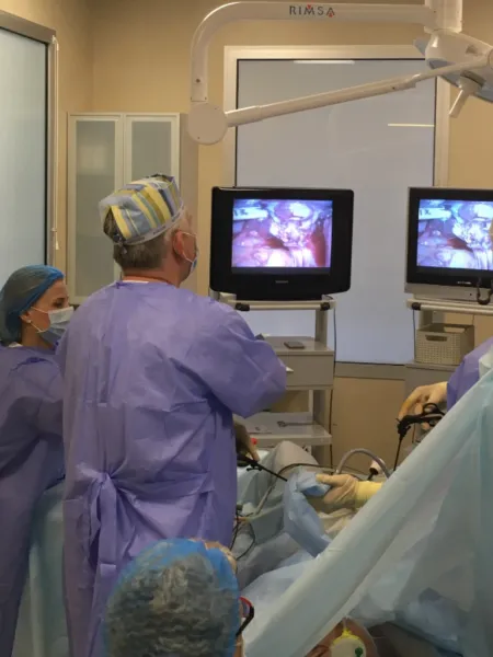
3. Examination of the mucous membrane under real-time video control.
4. If necessary, taking a sample for histological analysis.
After the procedure, the patient is observed for 1–2 hours and can go home the same day.
Advantages of Diagnostic Hysteroscopy
✅ Maximum accuracy — direct visual inspection of the uterine mucosa and structures
✅ Minimal trauma — no incisions or punctures
✅ Comfort and painlessness — performed under anesthesia
✅ Biopsy capability — diagnosis clarification in one intervention
✅ Outpatient procedure — does not require prolonged hospitalization
✅ Preparation for treatment — allows precise determination of therapy tactics or surgery planning
Preparation and Recovery
Preparation:
- Blood and urine tests, smears for flora, coagulogram
- Infection tests and ECG
- The procedure is usually conducted on the 6th–10th day of the menstrual cycle
After the procedure:
- Mild pulling pain and slight discharge for 1–3 days are normal
- Avoid sexual intercourse, visiting baths, pools, and physical exertion for 5–7 days
- Follow-up appointment with the doctor in 1–2 weeks
💡 Diagnostic hysteroscopy is an accurate, safe, and informative method for examining the uterine cavity, helping to identify hidden pathologies, clarify diagnoses, and choose the optimal treatment strategy. This procedure is indispensable in modern gynecology and reproductive medicine.
Hysteroscopy is a modern endoscopic method for examining and treating uterine cavity diseases, where the doctor inspects the inner surface of the uterus using a special instrument called a hysteroscope, which is inserted through the cervical canal without incisions or punctures.
In its diagnostic form, the procedure is used for precise visual assessment of the endometrium and uterine cavity, and if necessary, for taking a biopsy sample.
The method is considered the "gold standard" in gynecological diagnostics, surpassing ultrasound and other non-invasive studies in accuracy and informativeness.
When is Diagnostic Hysteroscopy Recommended
The procedure is recommended in the following cases:
- Menstrual cycle disorders (heavy, scanty, irregular periods)
- Uterine bleeding of unclear etiology
- Infertility and recurrent pregnancy loss
- Suspected polyps, fibroid nodules, or endometrial hyperplasia
- Follow-up after treatment or surgical intervention
- Clarification of ultrasound, hysterosalpingography, and other method results
- Preparation for IVF and other reproductive procedures
How the Procedure is Conducted
Diagnostic hysteroscopy is performed in an outpatient setting or hospital, most often under short-term intravenous anesthesia.
⏱ Duration of the procedure — 15–30 minutes.
Steps of the procedure:
1. Preparing the patient and administering light anesthesia.
2. Careful dilation of the cervical canal and insertion of the hysteroscope into the uterine cavity.
3. Examination of the mucous membrane under real-time video control.
4. If necessary, taking a sample for histological analysis.
After the procedure, the patient is observed for 1–2 hours and can go home the same day.
Advantages of Diagnostic Hysteroscopy
✅ Maximum accuracy — direct visual inspection of the uterine mucosa and structures
✅ Minimal trauma — no incisions or punctures
✅ Comfort and painlessness — performed under anesthesia
✅ Biopsy capability — diagnosis clarification in one intervention
✅ Outpatient procedure — does not require prolonged hospitalization
✅ Preparation for treatment — allows precise determination of therapy tactics or surgery planning
Preparation and Recovery
Preparation:
- Blood and urine tests, smears for flora, coagulogram
- Infection tests and ECG
- The procedure is usually conducted on the 6th–10th day of the menstrual cycle
After the procedure:
- Mild pulling pain and slight discharge for 1–3 days are normal
- Avoid sexual intercourse, visiting baths, pools, and physical exertion for 5–7 days
- Follow-up appointment with the doctor in 1–2 weeks
💡 Diagnostic hysteroscopy is an accurate, safe, and informative method for examining the uterine cavity, helping to identify hidden pathologies, clarify diagnoses, and choose the optimal treatment strategy. This procedure is indispensable in modern gynecology and reproductive medicine.


