In our clinic, we perform 4D fetal ultrasound, which allows you to clearly see the baby in three-dimensional space in motion.
Ultrasound for pregnant women helps diagnose abnormalities in the development of the fetus and the condition of the woman herself, provides a prognosis for the course of pregnancy and the timing of delivery. Additionally, it offers future parents the opportunity to meet thei...
In our clinic, we perform 4D fetal ultrasound, which allows you to clearly see the baby in three-dimensional space in motion.
Ultrasound for pregnant women helps diagnose abnormalities in the development of the fetus and the condition of the woman herself, provides a prognosis for the course of pregnancy and the timing of delivery. Additionally, it offers future parents the opportunity to meet their baby in advance and gain positive emotions from the pregnancy process.
- Experience with the most complex patients after IVF, as well as in cases of pregnancy loss;
- Involvement of related specialists when necessary;
- We conduct all types of research in prenatal diagnostics, as well as neonatal neurosonography;
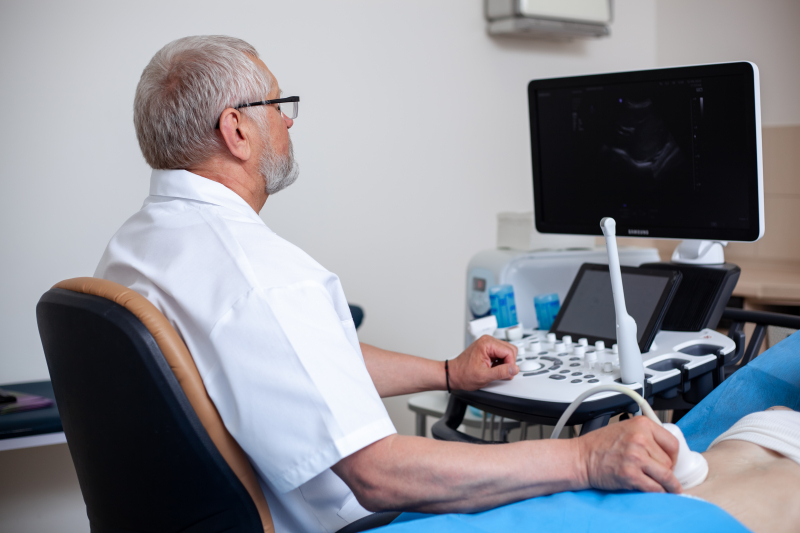
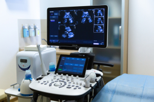
- Expert-class diagnostic equipment in a specialized ultrasound room -- SAMSUNG SH 50;
- Availability of Speckle-tracking echocardiography ...
- Experience with the most complex patients after IVF, as well as in cases of pregnancy loss;
- Involvement of related specialists when necessary;
- We conduct all types of research in prenatal diagnostics, as well as neonatal neurosonography;
- Expert-class diagnostic equipment in a specialized ultrasound room -- SAMSUNG SH 50;
- Availability of Speckle-tracking echocardiography function, allowing precise assessment of myocardial condition;
- Our sonologists are of the highest category with 20 years of experience;
- Video during the second and third trimester screenings -- as a gift.

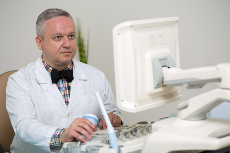
Lailo Dmitry Igorevich
Obstetrician-gynecologist, sonologist (ultrasound diagnostics doctor) of the highest category.
Fetal ultrasound - obstetric screening (I-II-III), pelvic ultrasound (gynecological ultrasound).

Lailo Valeria Viktorovna
Doctor-sonologist of the highest category
Ultrasound of internal organs


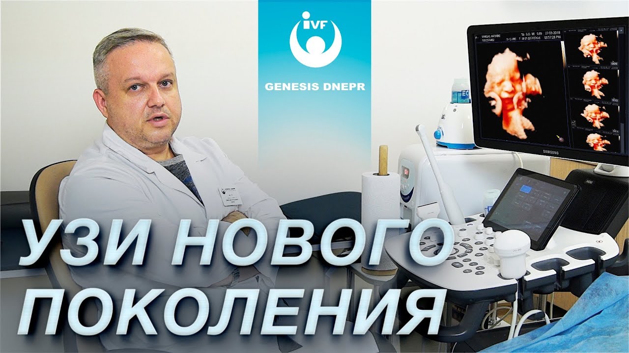
New generation ultrasound for pregnant women, obstetric screening at the "Genesis Dnipro" clinic. Doctor Lailo D.I.
Pregnancy ultrasounds are typically conducted at various stages of pregnancy to monitor fetal development and identify potential issues. This may include an initial ultrasound to determine the gestational age and assess fetal viability, as well as subsequent ultrasounds to evaluate the growth, weight, and development of the fetus.
Pregnancy ultrasound is a safe and non-invasive procedure that does not have harmful effects on the fetus or the uterus. It can be performed either externally (on the abdomen) or transvaginally (through the vagina), depending on the stage of pregnancy and the objectives of the examination.
Pregnancy ultrasound is an important part of prenatal medical care, allowing the doctor to monitor fetal development and promptly identify potential problems. This helps ensure the health and safety of both the mother and the fetus.
Fetal echocardiography is usually conducted in the second trimester of pregnancy and may be recommended in the following cases:
- If the woman has risk factors for developing heart pathologies in the fetus, such as genetic disorders or a family history of heart disease.
- If the doctor detected anomalies during previous ultrasounds and wants to examine the fetal heart in more detail.
- If the woman has symptoms or complaints that may indicate potential heart problems in the fetus.
Fetal echocardiography is performed using a specialized ultrasound machine that allows the doctor to obtain detailed images of the fetal heart. This may include assessing the heart's structure, chamber and valve sizes, valve function, blood supply, and other indicators that may be important for determining the heart's condition.
Fetal echocardiography is a safe procedure and does not have harmful effects on the fetus or the uterus. It can be performed either externally (on the abdomen) or transvaginally (through the vagina), depending on the stage of pregnancy and the objectives of the examination.
The results of fetal echocardiography can help the doctor make decisions about further treatment or monitoring of the fetus. This can be especially important when serious heart anomalies or pathologies are detected, which may require specialized medical care or early intervention.
An echocardiogram is performed using ultrasound waves, which are transmitted through the patient's body with a special device. The ultrasound waves reflect off various tissues and organs, and the collected data is processed by a computer to create a detailed image of the heart.
During an echocardiogram, the doctor can evaluate the heart's structure, the size of its chambers and valves, the function of the heart valves, blood supply, and other indicators that may be important for determining the heart's condition. This allows for the detection of possible abnormalities or pathologies, such as heart defects, arrhythmias, or other issues.
An echocardiogram may be prescribed in various situations, including checking the heart's condition in patients with symptoms or complaints, monitoring the heart's condition in patients with known heart diseases, and assessing the effectiveness of treatment and tracking changes in the heart.
This procedure is safe and non-invasive, meaning it does not require surgical intervention or injections. It can be performed externally (on the chest) or transvaginally (through the vagina), depending on the research goals and the patient's characteristics.
An echocardiogram is an important method for diagnosing and monitoring heart diseases. It allows the doctor to obtain detailed information about the heart's condition and take necessary measures to treat and improve the patient's health.
Neurosonography is performed using ultrasound waves transmitted through the infant's skull with a specialized device. These ultrasound waves reflect off various brain structures, and the data collected is processed by a computer to create a detailed image.
During neurosonography, a doctor can assess the brain's structure, the size of the brain's ventricles, the presence of hemorrhages or tumors, and other indicators that may be crucial for determining the brain's condition. This allows for the detection of possible abnormalities or pathologies, such as congenital brain defects or other issues.
Neurosonography may be recommended in various situations, including checking the brain condition of infants at risk of developing neurological problems, monitoring the brain condition of infants with known pathologies, and evaluating the effectiveness of treatment and tracking changes in the brain over time.
This procedure is safe and non-invasive for infants. It can be performed using a special transducer placed on the soft area of the infant's skull.
Neonatal neurosonography is an essential method for diagnosing and monitoring neurological issues. It provides doctors with detailed information about the brain's condition, enabling them to take necessary actions to treat and improve the child's health.
New generation ultrasound for pregnant women, obstetric screening at the "Genesis Dnipro" clinic. Doctor Lailo D.I.

Pregnancy support / management at the GENESIS DNEPR clinic. What does "all-inclusive" mean?

Birth with the Aroma of Coffee: How Private Maternity Hospitals Differ from Regular Ones. "Genesis Dnipro" Maternity Hospital
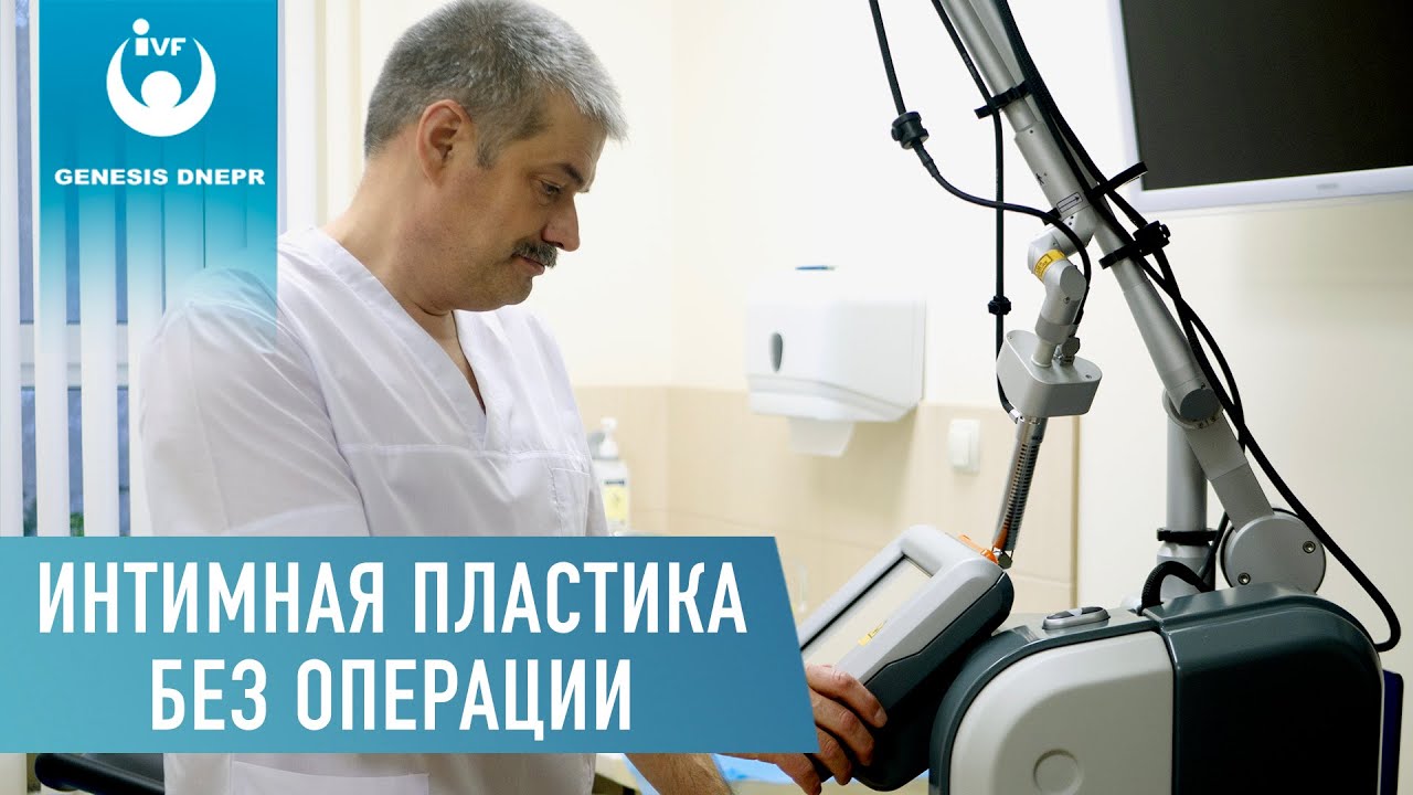
Intimate laser plastic surgery without pain and operation. Treatment of the intimate area with CO2 laser MonaLisa Touch Deka.

Tummy Tuck. How to Achieve a Beautiful Abdomen? Laser Plastic Surgery. Abdominoplasty | Surgeon Shevtsov
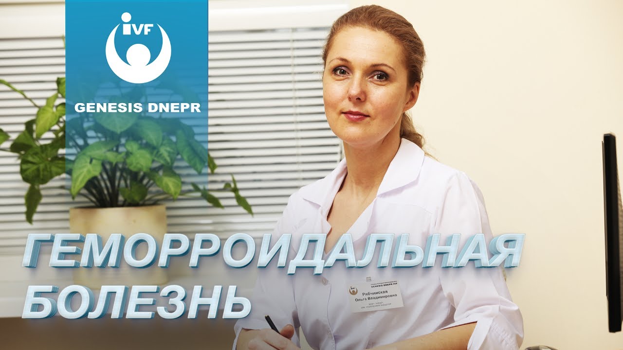
Hemorrhoidal disease. How to treat hemorrhoids. Proctologist Olga Vladimirovna Ryabchinskaya
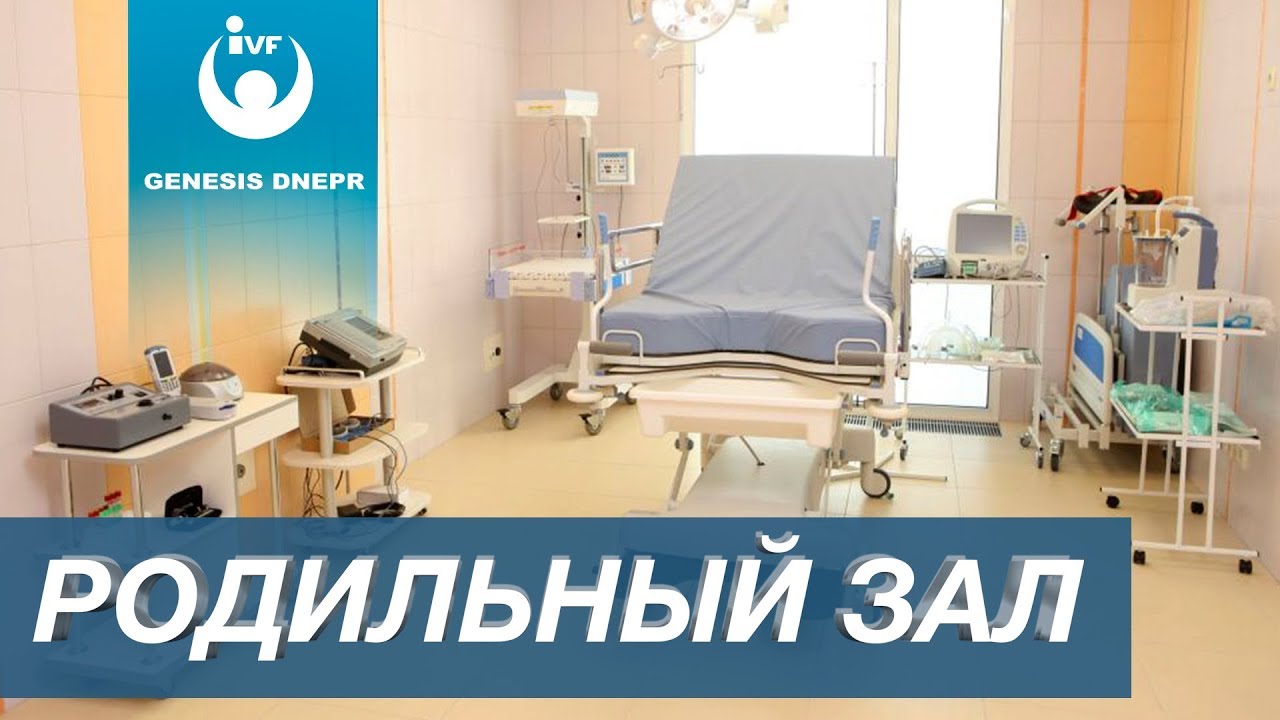
Genesis Dnepr Maternity Ward | Delivery, Room, Genesis Dnepr Maternity Hospital. How to Choose the Right Maternity Hospital.

About Genesis Dnepr Clinic / Genesis Dnepr Clinic

Cesarean Section: A Woman's Real Experience. Pregnancy and Childbirth at GENESIS DNEPR Clinic
1. ЗАПИС ПО ТЕЛЕФОНУ:
+38(067) 544-68-282. ЗАПИСЬ ЧЕРЕЗ АНКЕТУ:
Анкета слугує для попереднього знайомства лікаря з медичною інформацією про Вас та дає можливість записатися на платну чи безкоштовну консультацію ...
The clinic's reception phone number is 0963701324.
If you haven't made a decision yet and want to come for a tour, call the clinic's PR manager, Elena Ponomareva, at 0982412246.
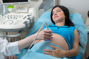
The ultrasound sensor is placed on the abdomen of the pregnant woman and is used to obtain images of the fetus and its movements. It operates based on the reflection of ultrasound waves from the tissues of the fetus and the uterus, allowing the doctor to see i...
The ultrasound sensor is placed on the abdomen of the pregnant woman and is used to obtain images of the fetus and its movements. It operates based on the reflection of ultrasound waves from the tissues of the fetus and the uterus, allowing the doctor to see its position, size, and other characteristics.
The Sonicaid fetal monitor allows the doctor to monitor the fetal heart rate and assess its condition. This can be particularly useful in cases where there are health risks to the fetus or when it is necessary to monitor its development.
However, it is important to note that the use of the Sonicaid fetal monitor should only be carried out under the supervision of a medical professional. Only they can interpret the monitoring results and take appropriate action if necessary.
долгота: 35.0320729


