In our clinic, we conduct the following heart and vascular s...
In our clinic, we conduct the following heart and vascular studies using ultrasound:
- Echocardiogram
- Doppler ultrasound of the head and limb vessels
- Fetal echocardiogram
- Neurosonography
We use cordyceps, Milan cocktail, and biopellets for effective treatment...
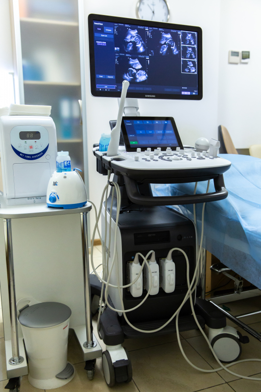
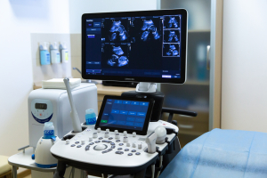
- Modern expert-class equipment, SAMSUNG SH 50;
- Availability of Speckle-tracking echocardiography function, which allows for accurate assessment of myocardial condition;

Voronezhskaya Tatyana Arkadyevna
Cardiologist of the highest category, rhythmologist.
Diagnosis and treatment of the cardiovascular system.

Mukhina Violetta Borisovna
Functional Diagnostics Physician. Heart ultrasound, ECG, Holter monitor.

Gilever Irina Andreevna
Ultrasound and functional diagnostics doctor. 17 years of experience.
Ultrasound of the abdominal organs, urinary system, prostate gland (including TRUS), scrotum, thyroid gland, breast, soft tissues, lungs; vessels of the head, neck, and lower extremities.


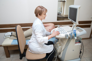

During an echocardiogram, the patient lies on their back while the doctor applies gel to the chest area and moves an ultrasound transducer over the chest. The transducer emits ultrasound waves that pass through the chest and reflect off the heart's structures, creating a detailed image on the screen.
An echocardiogram allows the doctor to evaluate several factors, including:
1. Heart function: The size and thickness of the heart walls, as well as the volumes and ejection fraction (the percentage of blood ejected from the ventricle with each heartbeat), are measured. This data helps determine how efficiently the heart is working and how its function can be improved.
2. Heart structures: The heart valves, atria, ventricles, and other structural features are assessed. This helps identify abnormalities such as valve narrowing or prolapse, and the presence of defects or anatomical disorders of the heart.
3. Heart movement: Ultrasound can be used to observe the movement of the heart walls and valves during contraction and relaxation (the cardiac cycle), which helps identify rhythm or coordination disorders of the heart's activity.
4. Blood flow: Echocardiography also allows for the assessment of the direction and extent of blood flow within the heart and vessels. The doctor can detect the presence of stenosis (narrowing) or regurgitation, which may indicate pathologies such as heart defects or valve problems.
An echocardiogram is a very useful tool for diagnosing and assessing the condition of the heart. It is highly accurate and is a virtually painless procedure. The results of the ultrasound examination help the doctor identify potential heart pathologies, tailor treatment, and monitor the effectiveness of therapy.
During a Doppler ultrasound, the patient typically sits or lies down while the doctor applies gel to the head and neck area. The doctor then uses an ultrasound probe (Doppler probe) to scan the relevant areas and conduct the examination.
The Doppler ultrasound of the head and neck vessels allows for:
1. Assessing blood flow: Sound waves help the doctor visualize blood flow in the arteries and veins of the head and neck. By analyzing the speed and direction of blood flow, the doctor can determine if there are any obstructions, narrowings, or constrictions in the vessels.
2. Detecting atherosclerosis: Atherosclerosis is a condition where cholesterol deposits, known as plaques, build up in the vessels. Doppler ultrasound can detect the presence of atherosclerosis by identifying vessel narrowing and changes in blood flow speed.
3. Diagnosing blood flow issues: Doppler ultrasound can also be used to identify blood flow problems, such as thrombosis (formation of blood clots in the vessels), embolism (movement of a clot or other particles in the blood), or stenosis (narrowing of a vessel).
4. Evaluating vascular structures: Doppler ultrasound can help the doctor obtain information about the structure of vessels, such as the arteries and veins of the head and neck. This can aid in identifying abnormalities or deformities in the vessels.
Doppler ultrasound of the head and neck vessels is a safe and non-invasive procedure. It provides high accuracy and helps the doctor obtain detailed information about the condition of the head and neck vessels. The results of this procedure can be used for further diagnosis and planning of treatment for relevant vascular issues.
A fetal echocardiogram is typically conducted between 20-22 weeks of pregnancy, although it can be performed at other times if necessary. The procedure involves applying gel to the mother's abdomen and using ultrasound technology to create an image of the fetal heart.
During a fetal echocardiogram, the doctor focuses on the following aspects:
1. Heart structure: The doctor evaluates various anatomical structures of the fetal heart, including valves, walls, and chambers. This helps identify potential anomalies and issues.
2. Heartbeat rhythm: The doctor determines the normal heart rhythm and checks for the presence of arrhythmias or irregularities.
3. Blood flow: The doctor analyzes the direction and speed of blood flow in the fetal heart to detect potential blood flow issues, such as stenosis or valve narrowing.
4. Other anomalies: A fetal echocardiogram can also help the doctor identify other anomalies related to the fetus, such as hydrocephalus or hydronephrosis.
This is an important procedure that helps in the early detection of potential fetal heart problems. As a result of the fetal echocardiogram, the doctor may take additional measures, including ordering further tests or consulting with a fetal cardiologist, to manage any identified problems or anomalies.
The procedure is performed using a special device called a neuro scanner, which is compact and can be easily moved around the baby's head. To conduct the examination, any hat or headband must be removed from the baby, and a gel is applied to the scalp to enhance the penetration of ultrasound waves.
During neurosonography, a specialist moves an ultrasound probe over the baby's scalp to obtain images of the brain. The examination typically takes about 15-20 minutes and does not cause discomfort to the baby.
Neurosonography may be recommended in the following cases:
- Suspected congenital brain anomalies;
- Presence of symptoms indicating possible brain function disorders (e.g., developmental delays, seizures, coordination issues);
- Premature birth or low birth weight;
- Presence of risk factors for brain pathologies (e.g., infections during pregnancy, genetic disorders in parents).
Neurosonography is a safe and non-invasive procedure that can be performed within the first few days of a baby's life. The results help the doctor assess the brain's condition and decide on the need for further examinations or treatment.
If brain pathologies are detected in a newborn, neurosonography may be repeated after a few months to monitor the disease's progression and the effectiveness of treatment.
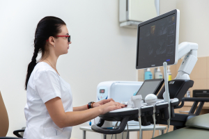
To perform STE, a standard ultrasound machine is used, but with additional software that enables tracking the movement of speckles on the heart's surface. During the examination, the patient lies on their back, and the doctor applies gel to the chest skin to enhance the penetration of ultrasound waves.
During STE, the doctor moves the ultrasound probe over the patient's chest skin and obtains images of the heart in both two-dimensional and three-dimensional modes. Then, using the software, the movement of speckles on the heart's surface is analyzed, allowing for the assessment of heart muscle function and the detection of abnormalities.
STE may be prescribed in the following cases:
- When there is a suspicion of heart function abnormalities;
- When there are risk factors for developing cardiovascular diseases (e.g., diabetes, hypertension, smoking);
- When monitoring the effectiveness of heart disease treatment.
STE is a safe and non-invasive procedure that can be performed at any age. The results of the examination help the doctor assess heart function and decide on the need for additional examinations or treatment.
In the case of detecting heart function abnormalities, STE can be repeated after a certain period to monitor the disease's progression and treatment effectiveness.
For DELV, an ultrasound machine with an additional sensor for measuring blood flow velocity is used. During the examination, the patient lies on their back, and the doctor applies gel to the skin to enhance the penetration of ultrasound waves.
During DELV, the doctor moves the sensor over the skin of the legs and obtains images of the vessels in both two-dimensional and color modes. Then, using software, the blood flow velocity in various vessels is analyzed.
DELV may be prescribed in the following cases:
- Suspected vascular diseases of the lower extremities (e.g., thrombosis, atherosclerosis);
- Presence of symptoms indicating circulatory disorders in the legs (pain, numbness, swelling);
- Monitoring the effectiveness of treatment for vascular diseases.
DELV is a safe and non-invasive procedure that can be performed at any age. The results of the examination help the doctor assess the condition of the leg vessels and decide on the need for additional examinations or treatment.
In cases where circulatory disorders in the legs are detected, DELV may be repeated after a certain period to monitor the disease's progression and the effectiveness of treatment.

Food intolerance. Food allergy. Examination. Therapist at Genesis Dnepr Clinic.
What is LIPOSUCTION? Laser liposuction, vibroliposuction, ultrasonic liposuction.|Surgeon Shevtsov

Lipofilling | Liposculpture of the face, neck, hands, and entire body. Mario Goisis and Igor Perelygin.

Treatment of Diabetes. Interview with an Endocrinologist.

The author of the weight loss book, I. V. Perelygin, talks about weight reduction | PART 1

Insulin resistance or excess weight. Treatment, symptoms, nutrition.
Bioidentical therapy, pellets, bioidentical hormones. Interview with Dr. Igor Perelygin.
How to prevent cardiovascular diseases? Dr. Gerasimenko Lyudmila at "Genesis Dnipro"

Who is eligible for plasmapheresis (blood purification) from viruses and diseases? Genesis Dnepr Clinic.
1. ЗАПИСЬ ПО ТЕЛЕФОНУ:
+38(067) 544-68-28+38 0963701324
- Heart ultrasound + speckle tracking echocardiography;
- ECG;
- Doppler ultrasound of the head vessels;
- Laboratory tests of blood and urine;
- Consultations with a cardiologist and a general practitioner.
For detailed information about the examination, please contact: +38 067 627 1905 - Nataliya Makarenko.
долгота: 35.0320729


