PROFESSOR, DOCTOR OF MEDICAL SCIENCES, ACADEMICIAN OF THE ACADEMY OF SCIENCES OF THE HIGHER SCHOOL OF UKRAINE
EXPERT IN OBSTETRICS AND GYNECOLOGY
NATURAL CHILDBIRTH
COMPLICATED CESAREAN SECTION WITH UTERINE SCAR, FIBROMYOMA, ENDOMETRIOSIS, AND UTERINE DEVELOPMENT ANOMALIES
ULTRASOUND DIAGNOSTICS OF ALL TYPES OF OBSTETRIC AND GYNECOLOGICAL PATHOLOGY
CONSERVATIVE MYOMECTOMY IN WOMEN WITH INFERTILITY
TREATMENT OF TRUE AND FUNCTIONAL OVARIAN CYSTS
...
PROFESSOR, DOCTOR OF MEDICAL SCIENCES, ACADEMICIAN OF THE ACADEMY OF SCIENCES OF THE HIGHER SCHOOL OF UKRAINE
EXPERT IN OBSTETRICS AND GYNECOLOGY
NATURAL CHILDBIRTH
COMPLICATED CESAREAN SECTION WITH UTERINE SCAR, FIBROMYOMA, ENDOMETRIOSIS, AND UTERINE DEVELOPMENT ANOMALIES
ULTRASOUND DIAGNOSTICS OF ALL TYPES OF OBSTETRIC AND GYNECOLOGICAL PATHOLOGY
CONSERVATIVE MYOMECTOMY IN WOMEN WITH INFERTILITY
TREATMENT OF TRUE AND FUNCTIONAL OVARIAN CYSTS
PUNCTURE OF FUNCTIONAL CYST
INFERTILITY
ABDOMINOCENTESIS IN CASE OF SUSPECTED FETAL DEVELOPMENT DISORDERS AND IN WOMEN OVER 40 YEARS OLD
Specialization: operative and general gynecology and obstetrics
OBSTETRICS:
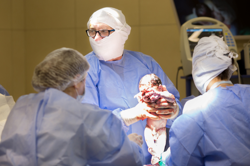
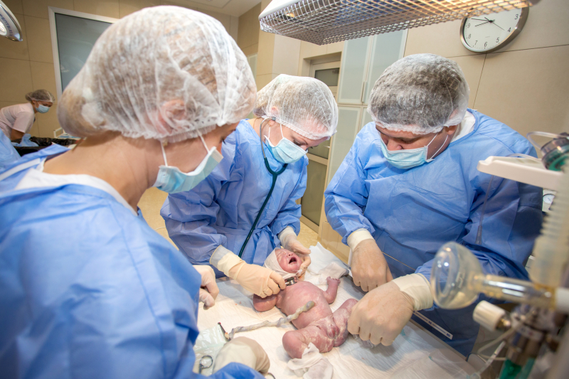
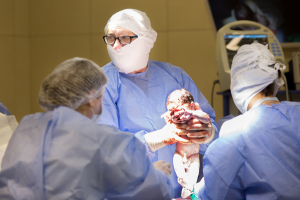
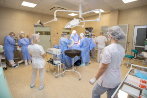
CESAREAN SECTION
performed in a modern operating room with "DRAGER" anesthesia equipment from Germany
CHILDBIRTH
conducted in a modern delivery room with a transformer bed for obstetric care
With subsequent patient stay in comfortable single rooms.
Secured parking, enhanced security measures, autonomous clinic operation.

Obstetrician-gynecologist, professor, Doctor of Medical Sciences, academician of the Academy of Sciences of the Higher School of Ukraine, honored worker of science and technology of Ukraine, head of the Department of Obstetrics and Gynecology at DGMU, chairman of the Dnipropetrovsk regional branch of the Association of Obstetricians and Gynecologists of Ukraine.
Surgery in obstetrics and gynecology, endocrine gynecology, reconstructive and restorative surgery in gynecology. Management of pregnancy with extragenital pathology.


In this case, the operation is complex due to the presence of a uterine scar, which may result from previous surgeries or childbirth, fibroids - tumors formed from uterine muscles, endometriosis - a condition where tissue similar to the inner lining of the uterus grows outside it, and uterine developmental anomalies - changes in the structure and shape of the uterus.
Such an operation requires special skills and experience from the surgeon to minimize risks and ensure the safety of both the mother and the child. Performing a cesarean section in such complex cases may require additional precautions and extra sutures to close the uterine wound.
Before the operation, the doctor carefully assesses the condition of the uterus, fibroids, endometriosis, and developmental anomalies to develop the most effective action plan. It is also important to discuss all possible risks with the patient to make an informed decision about proceeding with the surgery.
After the operation, the woman may require additional treatment or monitoring, especially in the presence of fibroids, endometriosis, or uterine developmental anomalies. The rehabilitation period after a complex cesarean section may be longer and requires special attention to wound healing and recovery of the body.
Ultrasound diagnostics is performed using a special device that emits ultrasound waves and receives echoes from organs and tissues. These echoes are then converted into an image on a screen, which can be analyzed by a doctor.
In obstetrics, ultrasound diagnostics is used to determine pregnancy, assess the development of the embryo or fetus, determine the baby's gender, detect any developmental abnormalities, and monitor the condition of the placenta. It can also be used to determine the causes of infertility, such as the presence of fibroids or polyps in the uterus, abnormalities in the structure of the ovaries, or fallopian tubes.
In gynecology, ultrasound diagnostics helps in identifying various diseases and pathologies, such as uterine fibroids, ovarian cysts, endometriosis, polyps, and tumors. It can also be used to monitor the condition of organs after surgery or treatment.
Ultrasound diagnostics is a safe and non-invasive procedure that does not require the use of radiation or the introduction of contrast agents. It can be performed at any phase of a woman's cycle and does not require special preparatory procedures.
However, to achieve the most accurate results, it is important that ultrasound diagnostics be performed by an experienced and qualified specialist. The doctor must have sufficient experience in interpreting the obtained data and identifying pathologies or changes.
Ultrasound diagnostics is an integral part of obstetric and gynecological practice. It allows doctors to obtain detailed information about the condition of organs and determine the presence of any pathologies or changes. This helps doctors make the right treatment decisions and improves outcomes for patients.
Uterine fibroids can be one of the factors preventing conception and successful pregnancy. The presence of fibroids in the uterus can cause a disruption in the anatomy of the uterine cavity, making it difficult for embryo implantation and pregnancy development. Additionally, fibroids can cause impaired blood flow in the uterus and increased muscle contractions, which also negatively affect pregnancy.
Conservative myomectomy allows for the removal of fibroids and the restoration of normal uterine anatomy. This can improve the chances of pregnancy and successful childbirth. However, it should be noted that the results of this procedure can be individual and depend on many factors, including the size and location of the fibroid, the woman's age, and the presence of other infertility factors.
Conservative myomectomy can be performed as an open surgical operation or using laparoscopic or robotic access. After the surgery, a woman may require a rehabilitation period for full recovery.
However, before performing a conservative myomectomy, a thorough examination and assessment of the condition of the uterus and other organs are necessary. Ultrasound diagnostics is an important method for determining the size and location of the fibroid, as well as for identifying other pathologies that may affect the outcome of the surgery.
Conservative myomectomy can be an effective method for treating uterine fibroids in women with infertility.
However, more active treatment may be necessary in some cases, particularly if the cyst is large, causes significant symptoms, or poses a health risk to the woman. In such cases, the following treatment methods may be recommended:
1. Surgical removal: This can involve laparoscopic or open surgery to remove the cyst. Laparoscopic surgery is a less invasive procedure performed through small incisions in the abdomen using special instruments. Open surgery requires a larger incision. Surgical removal may be recommended if the cyst is large, symptomatic, or poses a health risk.
2. Cyst drainage: This procedure involves removing fluid from the cyst using a needle and syringe. However, this method may be temporary, and the cyst may recur after drainage.
3. Hormonal therapy: In some cases, hormonal medications such as contraceptives or ovarian hormone antagonists may be recommended to help control cyst growth or prevent their occurrence.
4. Observation and monitoring: If the cyst is small and asymptomatic, the doctor may decide to simply monitor it and recheck its size and condition after some time.
During pregnancy, women may encounter various problems related to fetal development. These can include chromosomal abnormalities, genetic diseases, or other disorders that may affect the child's health and future. For women over 40, the risk of such problems increases, so a doctor may recommend abdominocentesis to obtain accurate information about the fetus's condition.
The abdominocentesis procedure is performed in a hospital or clinic setting under the guidance of an ultrasound scanner. Before the procedure, the patient may be offered a pain reliever or sedative to reduce discomfort.
During the procedure, the doctor uses an ultrasound scanner to determine the fetus's location and selects the safest spot for needle insertion. Then, using a special needle, the abdominal wall is punctured, and a small amount of fluid is extracted from the abdominal cavity. This fluid contains fetal cells that can be used for various tests.
After the procedure, the patient may be advised to remain under observation for a few hours to ensure there are no complications. Typically, after abdominocentesis, a woman can return to her normal life within a few days, although sometimes a longer recovery period may be necessary.
It is important to note that abdominocentesis is an invasive procedure and may carry risks of complications, such as infection or damage to the abdominal wall. Therefore, it is essential to thoroughly discuss all potential risks and contraindications with a doctor before undergoing the procedure.
Abdominocentesis is an important procedure for diagnosing fetal development disorders and making decisions about further treatment. However, it is crucial to thoroughly discuss all potential risks and contraindications with a doctor before proceeding. It is also important to monitor one's health and seek medical assistance for any changes in the body.
Functional ovarian cysts usually form as a result of normal ovulation processes and, in most cases, resolve on their own. However, sometimes they can grow large and cause discomfort or even pain. In such cases, a doctor may recommend aspiration to remove the cyst's contents and reduce its size.
The procedure for aspirating a functional cyst can be performed either in a hospital setting or at a clinic. Before the procedure, the patient may be offered pain relief or a sedative to reduce discomfort during the procedure.
During the aspiration, the doctor uses an ultrasound scanner to locate the cyst and selects the most convenient site for needle insertion. Then, using a special needle, the cyst is punctured, and its contents are aspirated. In some cases, a repeat aspiration may be necessary if the cyst is large or contains a significant amount of fluid.
After the procedure, the patient may be advised to remain under observation for a few hours to ensure there are no complications. Typically, after the aspiration of a functional cyst, a woman can return to her normal life within a few days, although sometimes a longer recovery period may be needed.
It is important to note that aspiration of a functional cyst is not a preventive method and does not prevent the formation of new cysts in the future. Therefore, it is important to consult a doctor if you experience pain or changes in your menstrual cycle to begin treatment promptly.
Aspiration of a functional ovarian cyst is an effective procedure for treating functional cysts and reducing their size. However, it is necessary to thoroughly discuss all possible risks and contraindications with a doctor before undergoing the procedure. It is also important to monitor your health and seek medical attention for any changes in your body.

A gynecologist answers the most common questions. Is emergency contraception dangerous?>

Doctors Perelygin I. V. and Shevtsov A. N. on plastic surgery at Genesis Dnepr clinic
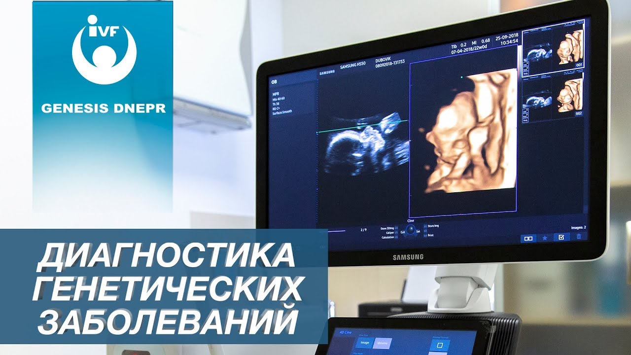
Diagnosis of Genetic Diseases in Future Children. Pregnancy and Childbirth. Professor V.A. Potapov.

Lipofilling | Liposculpture of the face, neck, hands, and entire body. Mario Goisis and Igor Perelygin
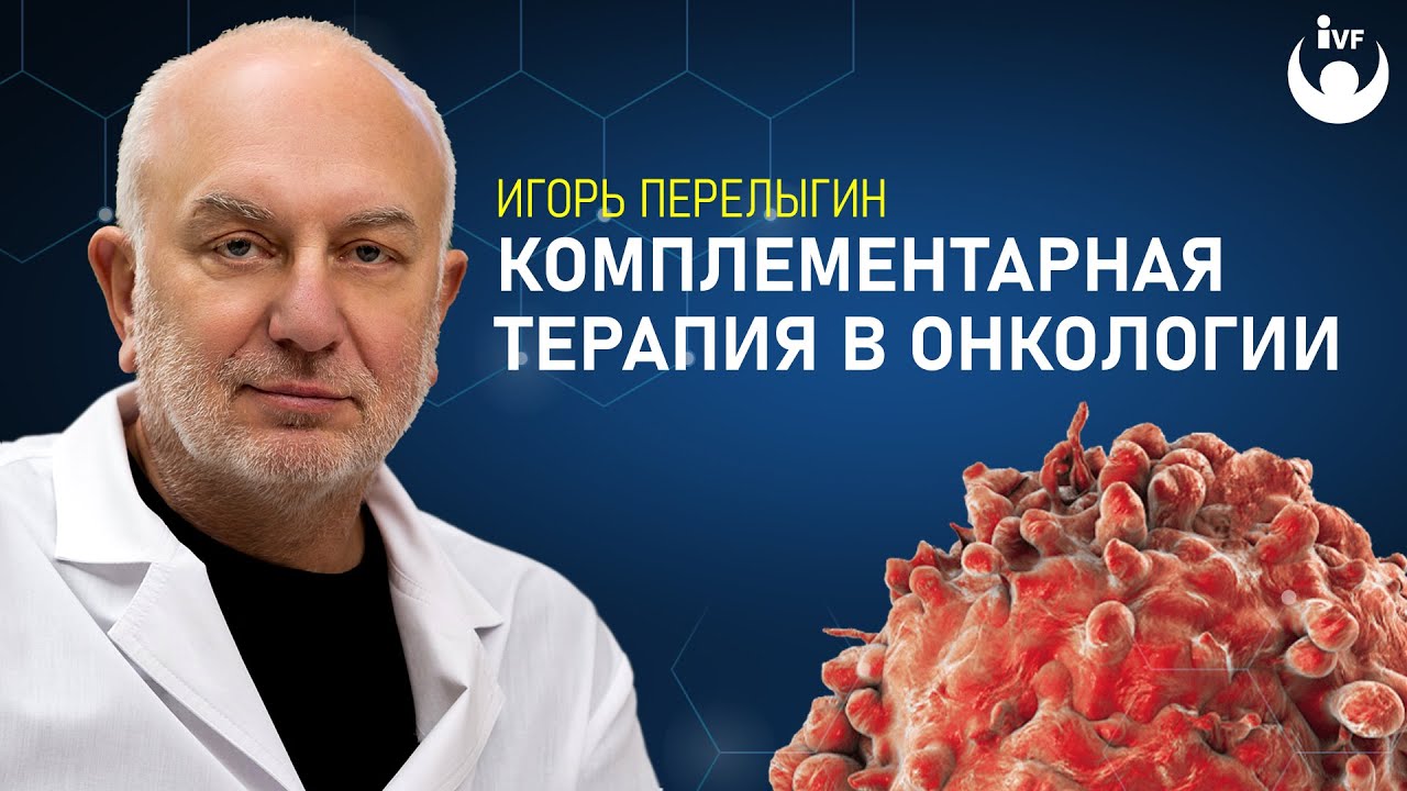
Alternative cancer treatment
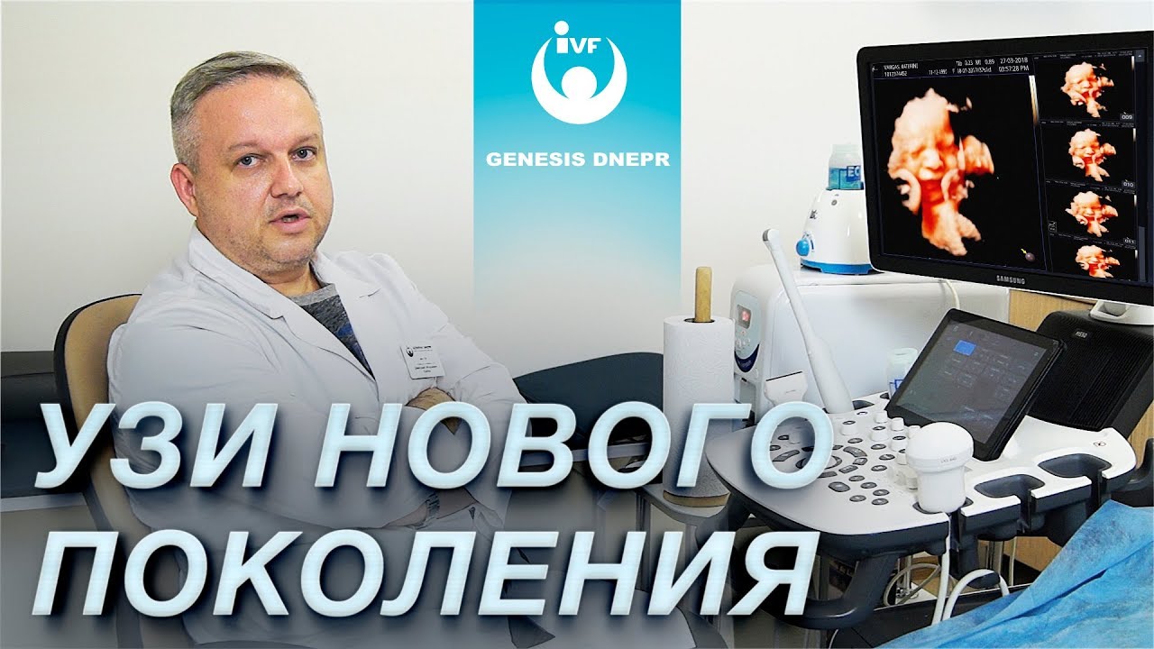
New generation ultrasound for pregnant women, obstetric screening at the "Genesis Dnipro" clinic.
долгота: 35.0320729


