This information is provided for reference purposes only and does not constitute advertising, an offer, a public offering, or a user manual. It is presented solely for the information of our patients abroad. This website does not engage in any form of distance selling. The sale, distribution, delivery, or prescription of products is not permitted through this website. Products and medical services may only be used as prescribed by a physician. Consultations are provided by clinic specialists in person or via remote medical consultations within the framework of current legislation. Please note that this directory does not programmatically permit the sale of products.
Ultrasound lung examination
Ultrasound of the lungs
Ultrasound examination of the lungs is a safe, painless, and highly informative diagnostic method that allows a doctor to assess the condition of the pleura, lung tissue, and surrounding structures. Although X-rays and CT scans remain the "gold standard" in diagnosing lung diseases, ultrasound is becoming increasingly popular due to its accessibility, speed, and lack of radiation exposure.
When Lung Ultrasound is Prescribed
The examination is conducted when there is suspicion of various respiratory system diseases, as well as for monitoring the condition after treatment or surgery. Indications for lung ultrasound include:
- Shortness of breath, cough, chest pain of unknown origin
- Suspected pneumonia or pleurisy
- Diagnosis of pleural effusion (fluid in the pleural cavity)
- Monitoring the dynamics of inflammatory processes
- Monitoring the condition after chest surgery
- Assessing the condition in patients with COVID-19 and other viral infections
What Lung Ultrasound Can Detect
Ultrasound examination provides the doctor with valuable information about the respiratory system's condition. It can help:
- Detect pleural effusion and determine its volume
- Identify inflammatory changes (e.g., pneumonia)
- Diagnose fibrotic changes and consolidations
- Assess the condition of the pleura and surrounding lung tissues
- Monitor the effectiveness of therapy
Advantages of the Method
✅ Safety — no radiation exposure, can be performed on children and pregnant women
✅ Painlessness and comfort — the examination does not cause discomfort
✅ High informativeness — especially for pathologies related to the pleura and superficial lung areas
✅ Possibility of dynamic observation — can be repeated as many times as necessary
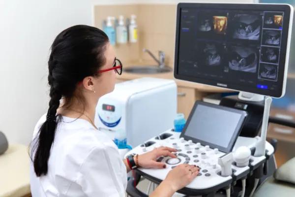
How the Procedure is Conducted
The patient takes a comfortable position sitting or lying down. The doctor applies a special gel to the skin and moves the transducer over the chest. The procedure takes no more than 10–15 minutes, requires no special preparation, and you can return to your usual activities immediately afterward.
💡 Lung ultrasound is a quick, accurate, and safe way to learn about the condition of your respiratory system. Regular examinations allow for the early detection of diseases and timely initiation of treatment.
When Lung Ultrasound is Prescribed
The examination is conducted when there is suspicion of various respiratory system diseases, as well as for monitoring the condition after treatment or surgery. Indications for lung ultrasound include:
- Shortness of breath, cough, chest pain of unknown origin
- Suspected pneumonia or pleurisy
- Diagnosis of pleural effusion (fluid in the pleural cavity)
- Monitoring the dynamics of inflammatory processes
- Monitoring the condition after chest surgery
- Assessing the condition in patients with COVID-19 and other viral infections
What Lung Ultrasound Can Detect
Ultrasound examination provides the doctor with valuable information about the respiratory system's condition. It can help:
- Detect pleural effusion and determine its volume
- Identify inflammatory changes (e.g., pneumonia)
- Diagnose fibrotic changes and consolidations
- Assess the condition of the pleura and surrounding lung tissues
- Monitor the effectiveness of therapy
Advantages of the Method
✅ Safety — no radiation exposure, can be performed on children and pregnant women
✅ Painlessness and comfort — the examination does not cause discomfort
✅ High informativeness — especially for pathologies related to the pleura and superficial lung areas
✅ Possibility of dynamic observation — can be repeated as many times as necessary
How the Procedure is Conducted
The patient takes a comfortable position sitting or lying down. The doctor applies a special gel to the skin and moves the transducer over the chest. The procedure takes no more than 10–15 minutes, requires no special preparation, and you can return to your usual activities immediately afterward.
💡 Lung ultrasound is a quick, accurate, and safe way to learn about the condition of your respiratory system. Regular examinations allow for the early detection of diseases and timely initiation of treatment.


