This information is provided for reference purposes only and does not constitute advertising, an offer, a public offering, or a user manual. It is presented solely for the information of our patients abroad. This website does not engage in any form of distance selling. The sale, distribution, delivery, or prescription of products is not permitted through this website. Products and medical services may only be used as prescribed by a physician. Consultations are provided by clinic specialists in person or via remote medical consultations within the framework of current legislation. Please note that this directory does not programmatically permit the sale of products.
Dopplerometry of the fetus
Dopplerometry during Pregnancy:
What is it?
• It is an ultrasound diagnostic method that allows for the assessment of blood flow in the vessels of the mother, placenta, and fetus.
• It is based on the Doppler effect, which is the change in frequency of ultrasound waves when reflected off moving particles (in this case, red blood cells).
• It is a safe and painless method that can be performed multiple times.
Why is Dopplerometry performed?
• To assess the delivery of oxygen and nutrients to the fetus through the placenta.
• To detect signs of hypoxia (oxygen deprivation of the baby).
• To diagnose intrauterine growth restriction of the fetus.
• To check the condition of the umbilical vessels, uterine arteries, and fetal brain vessels.
• To monitor the effectiveness of treatment in case of pregnancy complications.
What can be observed during the examination?
• The quality of blood flow between the mother, placenta, and fetus.
• Dysfunction of the placenta (placental insufficiency).
• Signs of hypoxia in the baby.
• Risk of fetal growth restriction.
When is it prescribed?
• Routinely, most often along with the third screening (30–34 weeks).
• Unscheduled, in cases of suspected hypoxia, oligohydramnios or polyhydramnios, fetal growth restriction, placental pathology, preeclampsia, or high blood pressure in the mother.
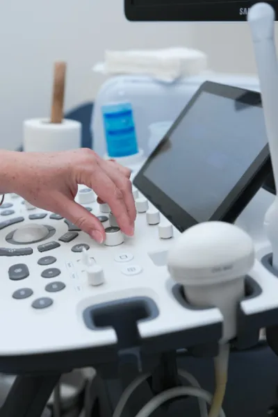
How is the procedure conducted?
• It is performed similarly to a regular ultrasound: the patient lies on a couch, and the doctor moves a transducer over the abdomen.
• Graphs and color images of blood flow in the vessels are displayed on the screen.
• The procedure takes about 10 minutes.
• No preparation is required.
What is it?
• It is an ultrasound diagnostic method that allows for the assessment of blood flow in the vessels of the mother, placenta, and fetus.
• It is based on the Doppler effect, which is the change in frequency of ultrasound waves when reflected off moving particles (in this case, red blood cells).
• It is a safe and painless method that can be performed multiple times.
Why is Dopplerometry performed?
• To assess the delivery of oxygen and nutrients to the fetus through the placenta.
• To detect signs of hypoxia (oxygen deprivation of the baby).
• To diagnose intrauterine growth restriction of the fetus.
• To check the condition of the umbilical vessels, uterine arteries, and fetal brain vessels.
• To monitor the effectiveness of treatment in case of pregnancy complications.
What can be observed during the examination?
• The quality of blood flow between the mother, placenta, and fetus.
• Dysfunction of the placenta (placental insufficiency).
• Signs of hypoxia in the baby.
• Risk of fetal growth restriction.
When is it prescribed?
• Routinely, most often along with the third screening (30–34 weeks).
• Unscheduled, in cases of suspected hypoxia, oligohydramnios or polyhydramnios, fetal growth restriction, placental pathology, preeclampsia, or high blood pressure in the mother.
How is the procedure conducted?
• It is performed similarly to a regular ultrasound: the patient lies on a couch, and the doctor moves a transducer over the abdomen.
• Graphs and color images of blood flow in the vessels are displayed on the screen.
• The procedure takes about 10 minutes.
• No preparation is required.


